1 Farrer Park Station Road, #12-03, Farrer Park Medical Centre, Singapore 217562 (65) 6443 7713 
Heel pain is a very common condition that affects patients in the 3rd to 5th decade of age. The most common conditions include plantar fasciitis as well as tendoachilles tendinitis. Plantar fasciitis involves inflammation of the plantar fascia which is a ligament in the heel bone. Due to overuse as well as trauma, it can result in aggravated symptoms of pain, difficulty in walking which is especially depicted in the early morning. As the condition worsens patient walking distance starts to decrease and they are unable to execute prolonged period of standing and walking.
Patients will present with heel pain, swelling as well as tight tendoachilles muscle structure in the posterior part of the calf which can result in severe calf pain. Following clinical assessment sometimes xray is require to evaluate the presence of unusual pathology such as bone cyst in the calcaneum as well as MRI to evaluate for any chronic injuries in the tendoachilles structure. In majority of patients the conditions can be managed with the following modalities which includes analgesia, supplements, weight reduction and appropriate advice on footwear orthotics and shoewear. The clinic physiotherapist will also treat the patient with ultrasound therapy, complimented by empowering the patient with muscle strengthening and exercises. However, in above 10 – 20% of patients due to their physical structure and work nature the condition maybe having limited response to medical management. As such these patients will then need to consider surgical intervention to improved the quality of life.
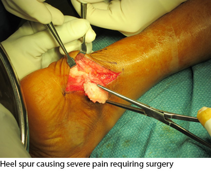
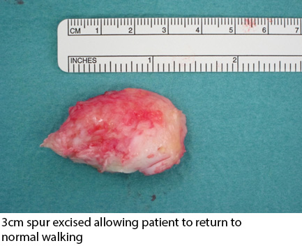
In these patients they may undergo the following procedures: i) release of the plantar fascia ligament; ii) PRP (Plasma Rich Precipitate) injection for chronic conditions; iii) gastrocnemius recession i.e. releasing the muscle tightness within the calf to reduce recurrence of plantar fasciitis symptoms; iv) in very aggravated tendoachilles tendinitis with retro calcaneum calcification they may benefit from surgical removal of these aggravating heel calcification which may cause recurrent tendoachilles tendinitis. Post-operatively therapy is very essential to prevent the relapse in such patients.
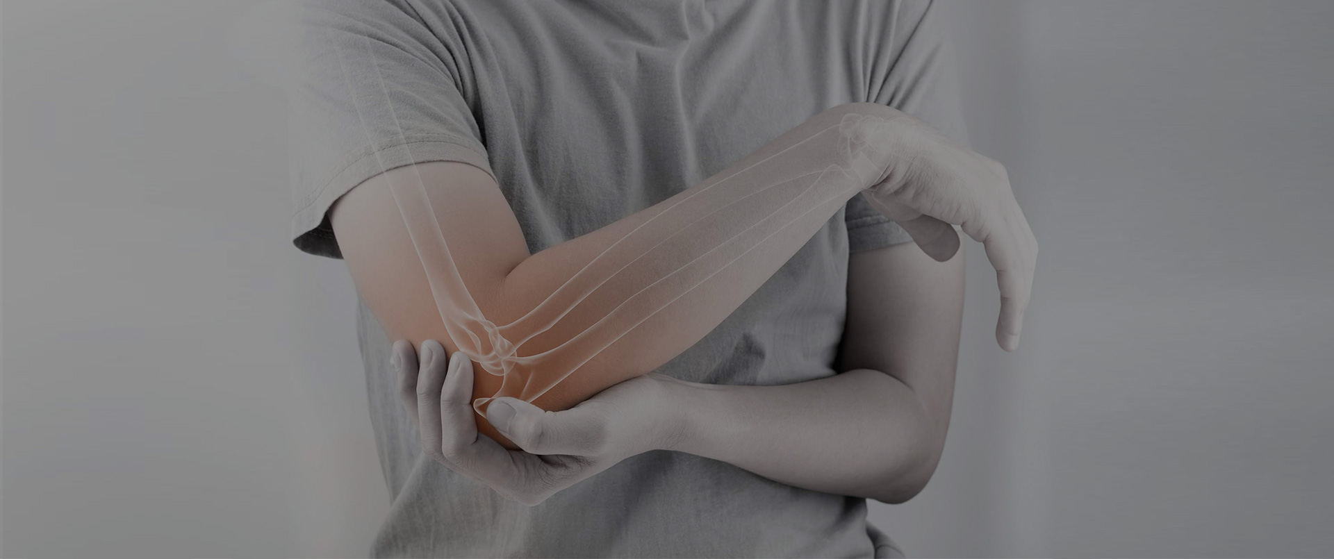
In this era where we are actively involve in usage of our upper limbs from multiples activities from heavy work load activities. Moreover, with continuous over use of computers and electronic equipment patients often present to the clinic with chronic elbow pain. Elbow pain can be in the form of either lateral epicondylitis (tennis elbow) or medial epicondylitis (golfer’s elbow) in patients they will need proper clinical assessment to evaluate the strength and the presentation of muscle weakness. Clinical evaluation with radiographs is sometimes necessary to ruled out the presents of calcification at the epicondyle.
In some patient the clinical assessment may need to be further assess for ruling out concomitant osteoarthritis of the elbow joint. In epicondylitis the management is usually non-surgical. They can be improved with off loading braces as well as appropriate physiotherapy. In persistent symptomatic cases they may benefit from clinic administration of cortisone injection. However, in severe recurrent cases they can benefit minimally invasive surgical techniques to release the adherent muscular attachment at the epicondyle. This is because the symptoms which are aggravated by chronic calcifications and result in chronic inflammation of the tendon attachments at either the medial epicondyle or lateral epicondyle. In patients who present with elbow arthritis they will however require different form of treatment which can be in the form of arthroscopic removal of loose bodies and removal of cartilage delaminations which will cause recurrent pain and swelling.
 Shoulder pain is a very common condition that affects patients in the middle ages. Recurrent pain and weakness can be due to rotator cuff tears. Rotator cuff tears in the small aspect can be managed with appropriate physiotherapy and cortisone injection.
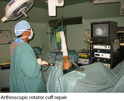
Large tears often result in patient presenting with either weakness or concomitant frozen shoulder. In such patient careful evaluation is required to facilitate return of the upper limb function with regards to activities of daily living and functional activities including strength for performing overhead arm movements. The rotator cuff is an essential muscle in keeping the humerus within the shoulder joint. The tear results in muscular efficiency reduction. Thus, they are having difficulty in performing overhead activities. In large tears these patients need to undergo arthroscopic repair of the muscle. Failure to do so results in weakness which will also be compounded by pain. With time the limited motion of the shoulder results in capsular scarring and the patient have frozen shoulder. These patient then present with limited function of the shoulder. By treating the patient early their clinical outcome is better and their return to activity is good.
Surgical treatment involves arthroscopy (keyhole) rotator cuff repair. By using dissolvable screws that are attached with sutures the muscle repair is delicately done and with augmented fixation techniques patients can return to physical strengthening modalities as fast as 2 to 3weeks following surgery. This enables early return to sporting activities and daily functional activities in most patient within 1 to 3months following surgical intervention depending on the severity of their condition.